

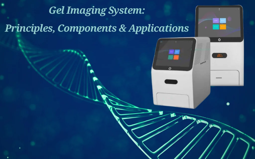
Introduction: Understanding the Gel Imaging System
A Gel Imaging System—also known as a gel documentation system or gel doc—is an essential laboratory instrument used to visualize, capture, and analyze results of gel electrophoresis and membrane blotting experiments. These systems are widely employed in molecular biology, biochemistry, and proteomics laboratories to detect, quantify, and document DNA, RNA, and protein bands stained with fluorescent, chemiluminescent, or colorimetric dyes.
Modern Gel Imaging Systems go beyond basic visualization, offering multi-spectral illumination, high-resolution digital imaging, and automated analysis software. This combination ensures precise, reproducible results and significantly enhances experimental efficiency.
What is a Gel Imaging System?
A gel imaging system is essentially a digital imaging platform designed to capture high-resolution images of gels and membranes. By combining illumination, detection, and software analysis, it allows researchers to measure molecular sizes, band intensity, and quantity with unparalleled accuracy.
Core Benefits:
High Sensitivity: Detect faint bands invisible to the naked eye
Quantitative Analysis: Accurately measure DNA, RNA, and protein concentrations
Safety: Blue light and LED illumination reduce UV exposure risks
Digital Documentation: Simplifies data sharing and long-term storage

How Does a Gel Imaging System Work?
The workflow of a gel imaging system typically includes:
Gel Preparation: Electrophoresis separates biomolecules based on size and charge.
Staining: Use dyes such as Ethidium Bromide, SYBR Safe, GelGreen, or Coomassie Blue.
Illumination: UV, blue, or LED light excites the dyes to emit detectable light.
Image Capture: A sensitive camera (CCD or CMOS) records the emitted light.
Analysis: Software quantifies bands, subtracts background, and exports data for publications or reporting.
")
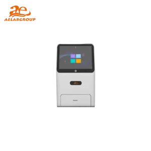
Core Components of a Gel Imaging System
1. Illumination Sources
UV Light (≈365 nm): Traditional method for DNA visualization; carries risk of DNA damage and user exposure.
Blue/Green Light (470–520 nm): Safer, compatible with non-toxic dyes such as SYBR Safe and GelGreen.
White Light: Ideal for protein gels stained with Coomassie or silver stains.
RGB Fluorescence: Detect multiple fluorophores in a single experiment.
Near-Infrared (NIR): Reduces autofluorescence and enhances sensitivity for low-abundance targets.
2. Detectors and Cameras
CCD Cameras: Low noise, high sensitivity, excellent for chemiluminescence.
CMOS Cameras: Fast, cost-effective, and increasingly comparable to CCDs.
Resolution: Ranges from 2 MP for basic tasks to 20 MP+ for high-precision quantitative imaging.
Field of View (FOV): Determines how much gel or membrane can be captured in one shot; larger FOVs increase workflow efficiency.
Detection Modes in Gel Imaging Systems
1. Fluorescence Imaging
Ideal for multiple fluorescent stains in a single gel. Excitation wavelengths match the dye to ensure optimal detection.
2. Chemiluminescence Detection
Gold standard for Western blot imaging, detecting faint protein bands without external excitation. Provides high dynamic range and reproducibility.
3. Visible Light Imaging
Used with colorimetric stains like Coomassie Blue or Ponceau S, essential for general protein quantification.
")
Software and Automation Features
Modern systems integrate advanced software platforms to streamline imaging and analysis:
Auto-Focus & Auto-Exposure: Ensures consistently sharp images.
Smart Lighting Selection: Automatically selects UV, blue, or white illumination.
Preset Protocols: Reduce repetitive tasks for consistent results.
Integrated Quantification Tools: Measure band intensity, background subtraction, and normalization.
Export & Sharing Options: Supports TIFF, JPEG, PDF, and data export for publications.
These features minimize user error and improve reproducibility in multi-user lab environments.
Field of View, Sensitivity, and Resolution
Field of View (FOV): Large FOVs allow imaging of multiple gels simultaneously, improving workflow.
Sensitivity: Critical for detecting faint bands or low-abundance proteins.
Resolution: High-resolution imaging (≥10 MP) ensures precise quantitative analysis and publication-quality data.
")
Safety and Eco-Friendly Alternatives
Traditional UV transilluminators can damage DNA and pose health risks. Blue/green light systems with non-toxic dyes provide safer and environmentally sustainable alternatives, supporting green lab initiatives worldwide. Compatible dyes include:
SYBR Safe
GelGreen
SeeGreen
SYBR Green
Applications of Gel Imaging Systems in Research
Gel imaging systems are indispensable across molecular biology, proteomics, and genomics:
DNA/RNA visualization and quantification
Western blot and protein analysis
2D gel electrophoresis
Colony counting and cell imaging
Multiplex fluorescence experiments
Immunoassays and binding studies
Post-translational modification research
This versatility ensures they remain central to modern research workflows.
")
Choosing the Right Gel Imaging System
Factors to Consider When Choosing the Right Gel Imaging System :
Detection Modes: Compatibility with fluorescence, chemiluminescence, or colorimetric stains.
Camera Resolution: ≥10 MP recommended for quantitative analysis.
Field of View: Ensure system accommodates your gel sizes.
Software Features: Automation, image analysis, and easy data export.
Safety: Prefer blue/green light illumination to reduce UV exposure.
Future-Proofing: Upgradeable systems for new fluorophores or detection technologies.

The Future of Gel Imaging Systems
The next generation integrates:
AI-powered quantification
Cloud connectivity
Faster image acquisition
Advanced LED and multi-spectral illumination
These innovations enable automated analysis, enhanced reproducibility, and improved research efficiency.
Conclusion: Why Modern Labs Need AELAB Gel Imaging Systems
A Gel Imaging System is no longer optional—it’s essential for precise, reproducible, and safe molecular research. From DNA visualization to protein quantification, these systems enhance workflow efficiency, accuracy, and lab safety.
Call to Action:
Upgrade your lab today with AELAB’s advanced Gel Imaging Systems. Explore our full range of solutions designed to improve electrophoresis documentation and ensure reliable, high-quality data for every experiment.