info@aelabgroup.com
QR Code 1
QR Code 2

Microscope Types and Their Applications in Detailed Study

Microscope Types and Their Applications in Detailed Study AELAB

Microscope Types and Their Applications in Detailed Study

Introduction to Microscopes

Microscopes are essential tools in scientific research and education, allowing researchers and students to observe objects that are too small to be seen with the naked eye. Understanding the different types of microscopes and their specific features is crucial for selecting the right one for a particular application. Whether you need high magnification, 3D imaging, or enhanced contrast, there’s a microscope suited for every need.

Types of Microscopes and Their Features

Ⅰ.Compound Microscopes


The compound microscope is one of the most commonly used types in laboratories and classrooms. It features multiple lenses—a primary objective lens and an ocular lens (eyepiece)—that work together to magnify specimens. This type of microscope is ideal for viewing thin, translucent specimens like cell slides. The combination of high magnification and resolution makes it suitable for studying biological samples.


Key Features:

  • Magnification: Typically ranges from 40x to 1000x.
  • Resolution: Can reveal details down to 200 nanometers.
  • Usage: Commonly used in biology, medical research, and education.

Ⅱ.Stereo Microscopes

Stereo microscopes, also known as dissecting microscopes, provide a three-dimensional view of specimens. Unlike compound microscopes, they use two separate optical paths to create a 3D image, making them ideal for examining larger, opaque specimens like insects, rocks, or electronic components.

Key Features:

  • Magnification: Generally between 10x and 70x.
  • Resolution: Not as high as compound microscopes but provides a detailed 3D view.
  • Usage: Suitable for dissection, assembly, and inspection tasks.

Ⅲ.Electron Microscopes


Electron microscopes use electron beams instead of light to achieve much higher magnifications and resolutions. There are two main types: Transmission Electron Microscopes (TEM) and Scanning Electron Microscopes (SEM).

Transmission Electron Microscopes (TEM): These microscopes transmit electrons through a very thin specimen, allowing for incredibly high magnification and detailed internal structure imaging.

  • Scanning Electron Microscopes (SEM): SEMs scan the surface of a specimen with electrons, producing detailed images of the surface topography.

Key Features:

  • Magnification: Can exceed 1,000,000x.
  • Resolution: Allows observation of structures at the atomic level.
  • Usage: Essential in materials science, nanotechnology, and detailed biological studies.

Ⅳ.Confocal Microscopes

Confocal microscopes use laser scanning and optical sectioning to create high-resolution images of thick specimens. By focusing on a single plane of the sample and excluding out-of-focus light, confocal microscopy provides sharper images and allows for 3D reconstruction.


Key Features:

  • Magnification: Generally from 10x to 100x.
  • Resolution: Higher than standard light microscopes, with better optical sectioning.
  • Usage: Commonly used in cellular biology and tissue imaging.

Ⅴ.Phase Contrast Microscopes

Phase contrast microscopes enhance the contrast of transparent specimens without staining. This is achieved by converting phase shifts of light passing through the specimen into variations in brightness, allowing for clear observation of live cells.


Key Features:

  • Magnification: Typically ranges from 10x to 100x.
  • Resolution: Similar to compound microscopes but with enhanced contrast.
  • Usage: Ideal for viewing live cells and other transparent specimens
Advanced Phase Contrast Microscope AELAB

Advanced Phase Contrast Microscope

Ⅵ.Polarizing Microscopes

Polarizing microscopes use polarized light to view materials that are anisotropic, meaning they have different properties in different directions. This technique is useful for studying minerals and crystals.


Key Features:

  • Magnification: Usually ranges from 10x to 100x.
  • Resolution: Similar to compound microscopes.
  • Usage: Primarily used in geology, mineralogy, and materials science.

Ⅶ.Digital Microscopes

Digital microscopes combine traditional microscopy with digital imaging technology. They often include a built-in camera and display, allowing for real-time observation and easy image capture and analysis.


Key Features:

  • Magnification: Varies widely depending on the model.
  • Resolution: Depends on the digital camera specifications.
  • Usage: Useful for educational purposes, quality control, and remote observation.

Conclusion

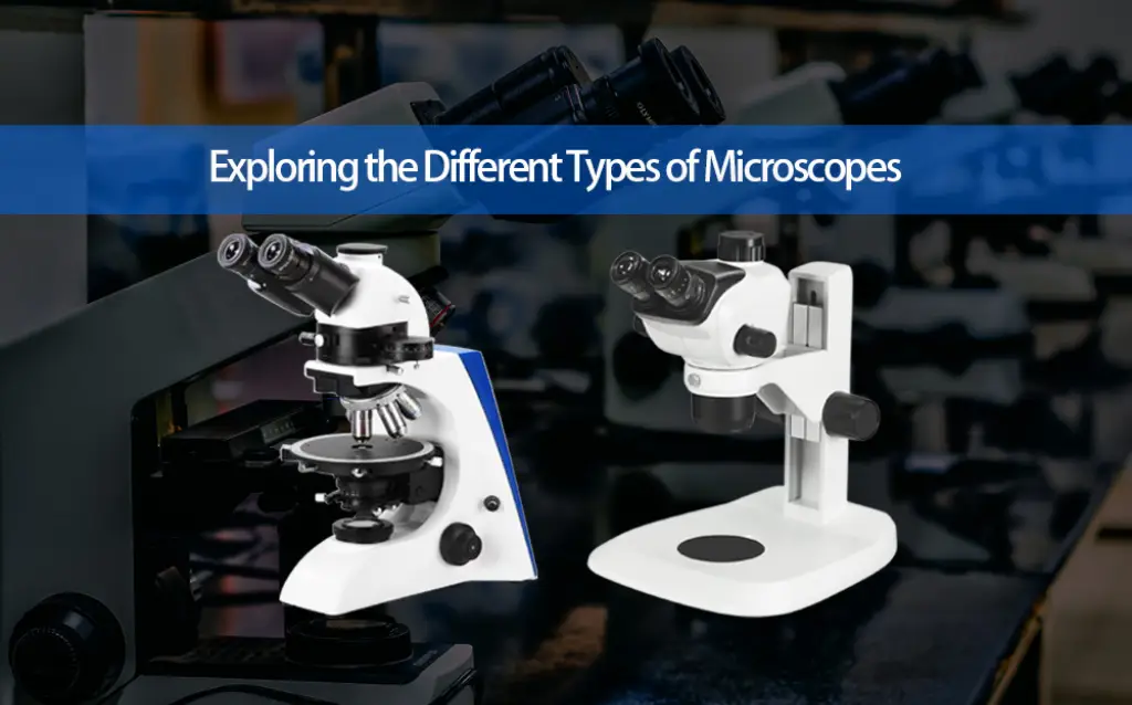
Microscopes are indispensable tools in scientific research, education, and industry. With various types such as compound microscopes, stereo microscopes, electron microscopes, and digital microscopes, each designed for specific tasks, understanding their features and capabilities is key to choosing the right one for your needs. Whether you are studying biological samples, inspecting materials, or conducting advanced research, the right microscope will provide the clarity and precision required for accurate results.

Frequently Asked Questions

1What is the main difference between a compound microscope and a stereo microscope?
A compound microscope uses multiple lenses to view thin, translucent samples at high magnification (up to 1000x), while a stereo microscope provides a lower-magnification (10x–70x) 3D view of larger, opaque objects like insects or circuit boards.
2How do electron microscopes achieve such high resolution?
Electron microscopes use electron beams instead of light, which have much shorter wavelengths. This allows them to magnify objects over a million times and visualize structures at the atomic level.
3Why is confocal microscopy especially useful for studying thick specimens?
Confocal microscopes use laser scanning and optical sectioning to focus on specific layers within thick samples, filtering out out-of-focus light and enabling high-resolution 3D imaging.
4In what situations would a phase contrast microscope be preferable?
Phase contrast microscopes are ideal for observing live, transparent cells without staining, as they enhance image contrast by converting light phase shifts into brightness differences.
5What makes polarizing microscopes essential in geology and materials science?
These microscopes use polarized light to analyze anisotropic materials—those with direction-dependent properties—making them crucial for studying minerals and crystal structures.
6How do digital microscopes benefit modern observation and education?
Digital microscopes integrate cameras and screens, allowing for real-time viewing, image capture, and analysis—making them especially useful for teaching, remote collaboration, and documentation.
Share:

Categories

Laboratory and Science Knowledge

Leave a Reply

Leave a Reply

Your email address will not be published. Required fields are marked *