

Microtome analysis plays a crucial role in biological research by enabling the precise sectioning of biological samples for detailed examination under a microscope. This technique is widely used in various fields, including histology, pathology, and forensic science. A microtome is an instrument designed to slice thin sections of tissue, allowing researchers to study the structure and function of cells and tissues with exceptional accuracy.
Applications of Microtome Analysis
Histology
Microtomes are fundamental in the preparation of tissue samples for histological examination. Thin tissue sections are essential for observing cellular structures and identifying disease patterns.
Pathology
Microtomes allow pathologists to examine tissue biopsies for diagnostic purposes, such as cancer detection. The ability to cut consistent, thin slices aids in accurate diagnosis and treatment planning.
Forensic Science
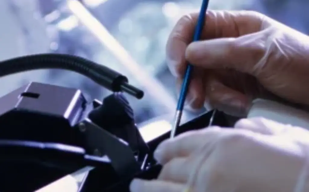
In forensic investigations, microtome analysis is used for analyzing paint chips, fibers, and other trace evidence. It helps in identifying the composition and layers of paint samples, which can link a suspect to a crime scene.
Neuroscience
Microtomes are essential for examining brain tissue. Studies on the neural structure and connectivity rely on high-precision sectioning to investigate complex neural pathways and tissue morphology.
Techniques Used in Microtome Analysis
Physical and Microscopic Examination
Researchers use microtomes to prepare samples for analysis under various types of microscopes, such as light, stereo, and polarized light microscopes. This allows them to examine the layers, thickness, and distribution of cellular components.
Frozen Sectioning
Technicians use a freezing microtome to section frozen tissue samples. This method is ideal for rapid diagnosis, particularly in surgical settings. They cryoprotect the tissue to minimize freezing artifacts, ensuring clear, high-quality sections.
Embedding and Thin-Sectioning
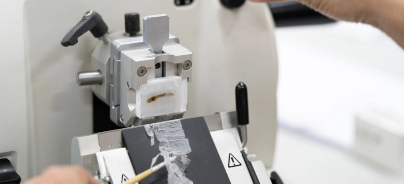
Researchers embed samples in a medium such as wax or epoxy resin to provide support during sectioning. They slice thin sections, typically ranging from 2 to 50 micrometers, using a sharp knife or diamond blade, preserving the tissue’s integrity for further analysis.
Polarized Light Microscopy
Scientists use polarized light microscopy (PLM) to examine the structural properties of paint films or other biological tissues. PLM effectively identifies specific components, such as pigments, additives, and contaminants, within the sample.
Types of Microtomes
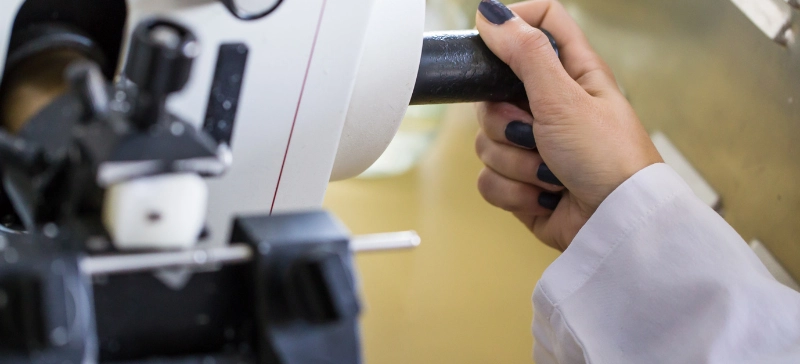
The rotary microtome is the most commonly used instrument for sectioning biological tissues. It rotates the tissue block against a stationary blade, producing precise, uniform sections. Users can adjust the section thickness by controlling the rotation speed.
Cryostat Microtome
Technicians house a cryostat in a cooling chamber, which allows them to section frozen tissue. This method is ideal for quickly processing tissues that need to stay at low temperatures to preserve their properties.
Vibratome
A vibratome uses a vibrating blade to section soft tissues, such as brain tissue, without the need for freezing. This type of microtome is particularly useful for maintaining tissue viability during experiments, especially in electrophysiological studies.
Sledge Microtome
The sledge microtome cuts thicker samples, typically embedded in harder materials like paraffin or synthetic resins. It uses a heavier, more robust blade to slice through tough tissue samples.
Ultramicrotome
Ultramicrotomes are specialized instruments used to cut extremely thin sections, usually for electron microscopy. They can produce sections as thin as 60 to 100 nanometers, providing high-resolution images of cellular structures.
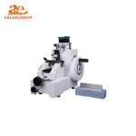
Aelab Microtome Products Overview
Aelab offers high-precision microtomes for accurate sectioning in biological, medical, and industrial applications. Their range includes:
- Rotary Microtome YD-1508R: Reliable with adjustable thickness (1-30 μm).
- Fully Automatic Microtome YD-355AT: Automated controls for sections (0.5–100 μm).
- Semi-Automatic Rotary Microtome YD-335A (Touch Screen): Easy operation with a 0.5-100 μm range.
- Semi-Automatic Rotary Microtome YD-335 (Button Panel): Precise control for routine histology.
- Rotary Microtome YD-315: For wax and plastic specimens (0-60 μm).
- Semi-Automatic Rotary Microtome YD-335A (LCD Screen): Adjustable thickness control (0.5-100 μm).
These microtomes provide precision, efficiency, and reproducibility for various laboratory applications. Each product is designed to enhance user efficiency and ensure precise, reproducible sections, making them ideal for various laboratory applications.

Conclusion
Microtome analysis is a vital tool in biological research, offering precise, reproducible sectioning for a wide range of applications. From histology to forensic science, microtomes enable researchers to examine tissue samples with unmatched accuracy. Understanding the different types of microtomes and their applications allows researchers to choose the most appropriate tool for their specific needs. With continuous advancements in microtome technology, this technique remains essential for exploring the complexities of biological systems at the cellular and molecular levels.
By mastering microtome analysis, researchers can unlock critical insights into tissue structures, diseases, and the underlying mechanisms of various biological processes.