

Introduction
The microscopic world reveals extraordinary details of tissues and cells that remain invisible to the naked eye. To explore these hidden structures, scientists prepare extremely thin and uniform slices of biological samples through a process called microtomy—the very foundation of histology and pathology. Among the essential tools for this technique, the Rotary Microtome plays a leading role in ensuring precise and reliable sectioning for advanced analysis.
")
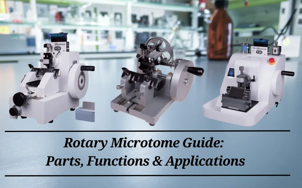
What is a Rotary Microtome?
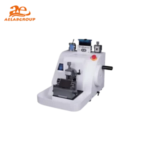
A rotary microtome is a precision mechanical device designed to cut thin sections of paraffin-embedded tissue for microscopic examination.
Working principle: A handwheel-driven mechanism moves the specimen block vertically against a stationary blade, producing slices ranging from 2–10 µm.
Etymology: The term microtome comes from the Greek mikros (small) and temnein (to cut).
Applications: Essential for preparing samples in light microscopy, immunohistochemistry, immunofluorescence, and electron microscopy.
Main Applications of Rotary Microtome
Rotary microtomes are versatile instruments widely used across different fields:
Clinical Pathology Labs: Dermatology, oncology, cardiology, ophthalmology, gynecology, and general diagnostic histology.
Universities and Research Centers: Studying tissue morphology and structural changes.
Pharmaceutical Companies & CROs: Used in drug development, genome sequencing, immunohistochemistry, and toxicology studies.
Teaching Laboratories & Service Providers: Training pathology students and outsourcing histology services.
")
Types of Microtomes
Different microtomes are available depending on applications:
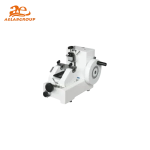
1. Rotary Microtome
Equipped with a single blade holder.
Allows continuous cutting.
Ideal for biopsies and serial sectioning.
2. Sliding Microtome
Blade moves horizontally across the specimen.
Suitable for large tissue blocks but less efficient for smaller samples.
3. Vibrating Microtome (Vibratome)
Cuts fresh, unfixed tissue.
Produces thicker slices (20–100 µm).
Widely used in neuroscience research.
4. Cryostat Microtome
Works at cryogenic temperatures (-20°C to -30°C).
Produces rapid frozen sections for intraoperative pathology.
")
Types of Rotary Microtomes
The devices are typically categorized into the following types
Manual Rotary Microtome: Handwheel-operated, cost-effective but less durable.
Semi-Automatic Rotary Microtome: Motor-assisted for greater accuracy.
Fully Automatic Rotary Microtome: Advanced models with programmable settings, ideal for research labs.
Key Components and Functions
Rotary microtomes consist of several essential parts:
Base & Housing: Provides stability and vibration-free operation.
Specimen Holder (Clamp): Secures tissue blocks in place.
Knife Holder: Holds disposable or reusable blades at adjustable angles.
Rotary Handwheel: Moves specimen block towards blade with controlled precision.
Thickness Adjustment Knob: Sets cutting thickness (0.25–100 µm depending on model).
Injection System: Ensures section thickness accuracy down to 0.5 µm.
Safety Features: Dual-lock handwheel and retraction system for operator safety.
")
Rotary Microtome Maintenance
Proper care extends lifespan and ensures accuracy:
Annual Inspection: Professional service check.
Cleaning: Use distilled water, avoid harsh chemicals.
Mechanical Care: Tighten screws, lubricate parts, check vibrations.
Record Keeping: Track part replacements and maintenance history.
Storage: Cover and disconnect when not in use.
 (1)")
10 Professional Tips for Better Section Quality
Keep blades sharp and replace regularly.
Adjust cutting speed depending on tissue hardness.
Keep samples cool to reduce compression artifacts.
Ensure microtome table is perfectly level.
Clean and calibrate equipment frequently.
Fix samples firmly to avoid movement.
Use proper embedding media.
Avoid prolonged continuous sectioning.
Monitor handwheel ergonomics to prevent operator strain.
Maintain detailed records of adjustments and performance.
Advantages
- High precision and stability.
- Adjustable cutting angles.
- Suitable for all tissue types (soft, hard, fatty).
- Produces continuous ribbons.
- Reduced vibration during sectioning.
- Compatible with advanced lab automation.
")
Rotary Microtome vs Other Microtomes
| Feature | Rotary Microtome | Cryostat Microtome | Vibrating Microtome | Sliding Microtome |
|---|---|---|---|---|
| Cutting Medium | Paraffin-embedded tissues | Frozen tissues | Fresh tissues | Large blocks |
| Section Thickness | 1–10 µm | 4–20 µm | 20–100 µm | 10–60 µm |
| Applications | Histopathology, biopsies | Rapid diagnosis | Neuroscience | Research on large samples |
| Speed | Moderate | Very fast | Slow | Moderate |
Conclusion
The rotary microtome is an indispensable instrument in histology, pathology, and biomedical research. By enabling precise and reproducible tissue sectioning, it bridges the microscopic world with diagnostic and research needs.
Choosing the right type—manual, semi-automatic, or fully automatic—along with proper training, regular maintenance, and adherence to professional sectioning techniques ensures consistent high-quality results.
👉 Call to Action: If you’re a lab professional or researcher, consider investing in a high-quality rotary microtome from AELAB to enhance accuracy, efficiency, and reliability in your diagnostic and research work.