AELAB | Analytical Equipment | X-Ray Diffractometer (XRD)
The Laboratory XRD Spectrometer (X-Ray Diffraction Spectrometer) is a cornerstone instrument in material characterization, enabling precise analysis of crystal structures, phase composition, and molecular arrangements. By measuring how X-rays diffract through a material’s lattice, XRD provides crucial data for identifying compounds and understanding material properties. Commonly used in materials science, chemistry, pharmaceuticals, and geology, Laboratory XRD Spectrometers deliver non-destructive, high-resolution insights into crystalline substances ranging from minerals and metals to polymers and pharmaceuticals. Their versatility and precision make them indispensable in both research and quality control environments, supporting advancements in nanotechnology, manufacturing, and advanced materials development.
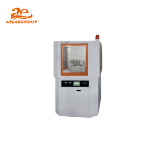
A Laboratory XRD Spectrometer is an analytical device used to determine the crystallographic structure of materials by analyzing the diffraction of X-rays. When a beam of X-rays strikes a crystalline sample, the rays are diffracted according to the atomic arrangement within the material. The instrument records these diffraction patterns and translates them into data that reveal the crystal phase, lattice spacing, and degree of crystallinity. XRD spectrometers are fundamental for identifying unknown compounds, verifying structural integrity, and studying phase transitions in metals, ceramics, polymers, and pharmaceuticals.
| Feature | Specification |
|---|---|
| X-Ray Source | Cu Kα radiation (1.5406 Å), with Mo or Cr optional |
| Operating Voltage | 20–60 kV adjustable, depending on tube type |
| Detector Type | Scintillation or CCD detector for high sensitivity |
| Angular Range | 5°–90° 2θ typical, extendable for advanced analysis |
| Resolution | High, capable of distinguishing closely spaced peaks |
| Software Integration | Automated phase ID, Rietveld refinement, database comparison |
| Sample Compatibility | Powders, thin films, solids, nanomaterials |
| Compliance | Meets ISO and ASTM standards for diffraction analysis |
| Aspect | XRD Spectrometer | XRF Analyzer | SEM Microscope |
|---|---|---|---|
| Principle | X-ray diffraction by crystal lattice | X-ray fluorescence of elements | Electron-sample surface interaction |
| Main Output | Crystal structure and phase composition | Elemental composition | Surface morphology and imaging |
| Sample Preparation | Minimal, typically fine powder | Minimal | Requires conductive coating |
| Resolution | High for crystal structure | Moderate to high for elements | High for topography |
| Analysis Speed | Moderate (minutes) | Fast (seconds) | Moderate to slow |
Q: What information can XRD provide about a sample?
A: XRD reveals the crystal structure, phase composition, crystallinity, and lattice parameters of a material, enabling precise identification of compounds and structural changes.
Q: Can XRD analyze amorphous materials?
A: No, XRD is suitable only for crystalline materials. Amorphous substances produce broad, diffuse scattering rather than distinct diffraction peaks.
Q: What sample size is required for XRD analysis?
A: Typically, a few milligrams of finely ground sample are sufficient for accurate diffraction measurement.
Q: How long does a typical XRD scan take?
A: Depending on the resolution and angular range, a scan may take from a few minutes to about an hour for high-precision measurements.
Laboratory XRD Spectrometers adhere to international standards such as ISO and ASTM for diffraction analysis and phase identification. AELAB supports full calibration, certification, and documentation services to ensure compliance and optimal instrument performance in research and industrial laboratories.
Looking for specific lab equipment? Fill out the form below, and our team will get back to you with detailed information and a personalized quote.