Comprehensive Guide to Laboratory Biological Microscopes
Introduction
Understanding the intricate details of biological organisms requires advanced tools, among which laboratory biological microscopes stand out prominently. These precision instruments are indispensable for scientific research, medical diagnostics, and education. In this guide, we’ll explore their key components, technical specifications, advantages, limitations, and expert recommendations to help you choose and use them effectively.
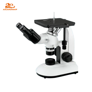
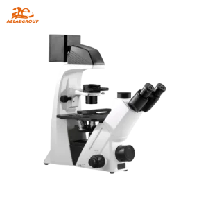
What Is a Laboratory Biological Microscope?
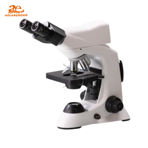
A laboratory biological microscope is designed to magnify and visualize biological samples such as cells, tissues, and microorganisms that are invisible to the naked eye. By using advanced optical systems and illumination technologies, it provides clear, detailed 2D images crucial for research and diagnostics.
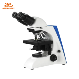
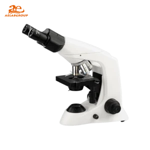
Key Components
- Eyepiece (Ocular Lens): Provides 10x magnification for observation.
- Objective Lenses: Offer various magnifications—typically 4x, 10x, 40x, and 100x (oil immersion).
- Stage: Holds specimen slides securely for precise positioning.
- Illumination System: Ensures consistent lighting using LED or halogen sources.
- Focusing Mechanism: Coarse and fine focus knobs allow sharp, clear images.
Detailed Technical Features
| Feature |
Details |
| Magnification Range |
Typically 40× to 1000× (up to 2000× in advanced models) |
| Optical System |
Achromatic or plan-achromatic lenses for high clarity |
| Illumination Type |
LED (cool, energy-efficient) or halogen (intense, warm light) |
| Focus Mechanism |
Coarse and fine adjustment knobs with smooth gearing |
| Stage Type |
Mechanical stage with slide holder and XY controls |
| Head Configuration |
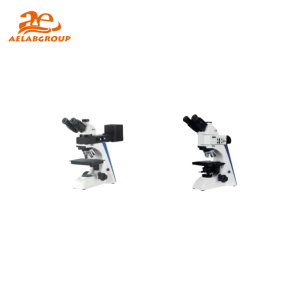
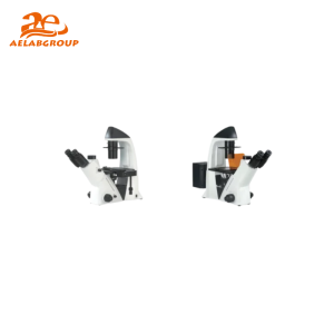
Monocular, binocular, or trinocular (for digital cameras) |
| Digital Integration |
USB or Wi-Fi camera connectivity for image capture and analysis |
| Power Supply |
AC or rechargeable battery-powered options available |
Benefits of Laboratory Biological Microscopes
- Enhanced Precision: Enables detailed and reliable microscopic observations.
- Versatile Applications: Used in research, diagnostics, education, and industrial biology.
- Educational Value: Ideal for teaching fundamental biological concepts and microscopy techniques.
- Diagnostic Utility: Supports pathology, microbiology, and cytology by identifying microorganisms and tissue abnormalities.
Limitations and Considerations
- Cost: High-end models with digital or research-grade optics can be expensive.
- Maintenance: Regular cleaning, calibration, and light source checks are essential.
- Training: Users need basic microscopy training for accurate and safe operation.
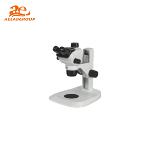
Biological Microscope vs. Stereo Microscope
| Aspect |
Biological Microscope |
Stereo Microscope |
| Image Type |
2D, high-magnification image |
3D image with depth perception |
| Magnification Range |
40× to 1000× |
7× to 90× |
| Best For |
Cells, bacteria, and tissues |
Insects, plants, and small mechanical parts |
| Lighting |
Transmitted light through the specimen |
Reflected light from specimen surface |
| Image Depth |
Flat, highly magnified detail |
Depth-rich, surface-level view |
Professional Tips for Maximizing Microscope Performance
- Prepare slides carefully using clean coverslips to prevent contamination.
- Perform calibration routinely to ensure consistent magnification accuracy.
- Use fine adjustment for focusing at high magnification to avoid damaging slides.
- Integrate a digital camera system for documentation and data sharing.
- Keep optics dust-free and handle with care to prolong the microscope’s lifespan.
How to Choose the Right Laboratory Biological Microscope
- Define Your Application: Identify whether you’ll study cells, tissues, or microorganisms.
- Check Optical Quality: Look for achromatic or plan-achromatic objectives for clarity.
- Evaluate Illumination Type: Choose LED for energy efficiency or halogen for intensity.
- Determine Magnification Needs: Ensure the model supports your desired magnification range.
- Balance Budget and Quality: Select a reliable model that fits your requirements and budget.
Maintenance and Best Practices
- Clean lenses regularly using lens paper and approved cleaning solutions.
- Cover the microscope when not in use to prevent dust accumulation.
- Check and align illumination systems periodically.
- Lubricate mechanical parts as recommended by the manufacturer.
- Have the microscope professionally serviced annually for calibration and alignment.
FAQ
Q: What magnification is best for observing bacteria?
A: A 100× oil immersion objective combined with a 10× eyepiece (1000× total) provides the clarity needed for bacterial observation.
Q: Can I attach a camera to a biological microscope?
A: Yes. Trinocular models are designed for camera integration, allowing high-resolution image and video capture.
Q: How often should a biological microscope be calibrated?
A: Calibration should be performed every 6–12 months, or more frequently for high-precision research applications.
Q: Are LED microscopes better than halogen ones?
A: LED microscopes are generally preferred for their low heat output, consistent brightness, and energy efficiency.