AELAB | Laboratory Equipment | Microscope | Biological Microscope
Understanding the intricate details of biological organisms requires advanced tools, among which laboratory biological microscopes stand out prominently. These precision instruments are indispensable for scientific research, medical diagnostics, and education. In this guide, we’ll explore their key components, technical specifications, advantages, limitations, and expert recommendations to help you choose and use them effectively.
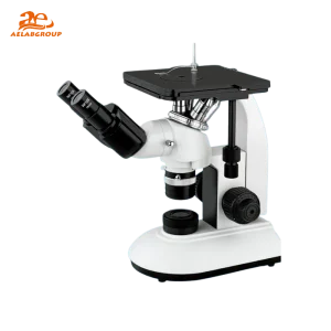
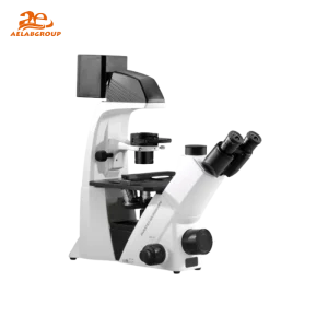
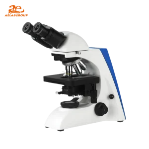
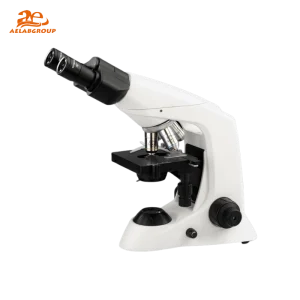
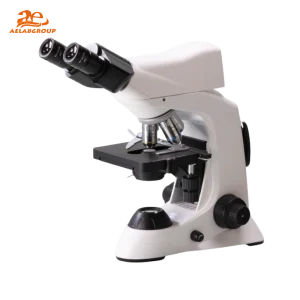
A laboratory biological microscope is designed to magnify and visualize biological samples such as cells, tissues, and microorganisms that are invisible to the naked eye. By using advanced optical systems and illumination technologies, it provides clear, detailed 2D images crucial for research and diagnostics.
| Feature | Details |
|---|---|
| Magnification Range | Typically 40× to 1000× (up to 2000× in advanced models) |
| Optical System | Achromatic or plan-achromatic lenses for high clarity |
| Illumination Type | LED (cool, energy-efficient) or halogen (intense, warm light) |
| Focus Mechanism | Coarse and fine adjustment knobs with smooth gearing |
| Stage Type | Mechanical stage with slide holder and XY controls |
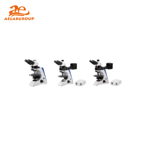
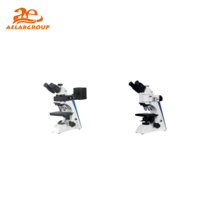
| Head Configuration | Monocular, binocular, or trinocular (for digital cameras) |
| Digital Integration | USB or Wi-Fi camera connectivity for image capture and analysis |
| Power Supply | AC or rechargeable battery-powered options available |
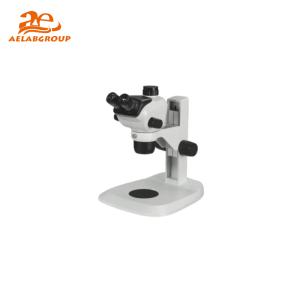
| Aspect | Biological Microscope | Stereo Microscope |
|---|---|---|
| Image Type | 2D, high-magnification image | 3D image with depth perception |
| Magnification Range | 40× to 1000× | 7× to 90× |
| Best For | Cells, bacteria, and tissues | Insects, plants, and small mechanical parts |
| Lighting | Transmitted light through the specimen | Reflected light from specimen surface |
| Image Depth | Flat, highly magnified detail | Depth-rich, surface-level view |
Q: What magnification is best for observing bacteria?
A: A 100× oil immersion objective combined with a 10× eyepiece (1000× total) provides the clarity needed for bacterial observation.
Q: Can I attach a camera to a biological microscope?
A: Yes. Trinocular models are designed for camera integration, allowing high-resolution image and video capture.
Q: How often should a biological microscope be calibrated?
A: Calibration should be performed every 6–12 months, or more frequently for high-precision research applications.
Q: Are LED microscopes better than halogen ones?
A: LED microscopes are generally preferred for their low heat output, consistent brightness, and energy efficiency.
Looking for specific lab equipment? Fill out the form below, and our team will get back to you with detailed information and a personalized quote.