AELAB | Laboratory Equipment | Microscope | Fluorescence Microscope
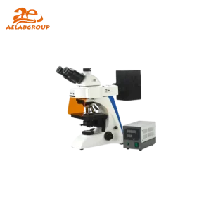
A laboratory fluorescence microscope is an advanced optical instrument that uses fluorescence to visualize and analyze biological, chemical, and material samples with exceptional contrast and specificity. By detecting light emitted from fluorescent dyes or proteins, it enables researchers to study cellular structures, molecular interactions, and biochemical processes in unprecedented detail. This technology has become a cornerstone of modern life science and diagnostic laboratories.
A fluorescence microscope is a specialized type of optical microscope that illuminates samples with specific wavelengths of light to excite fluorescent molecules. These molecules then emit light at a longer wavelength, which is detected and filtered to produce high-contrast images. This selective illumination allows visualization of specific components within cells or materials, providing detailed insights into biological and chemical phenomena.
| Feature | Details |
|---|---|
| Light Source | High-intensity mercury, xenon, or LED lamps for excitation |
| Excitation & Emission Filters | Precisely control wavelength ranges for specific fluorophores |
| Detection System | High-sensitivity cameras (CCD or CMOS) and photomultiplier detectors |
| Fluorescence Channels | Supports multiple markers for multi-label imaging |
| Magnification | Typically 40×–1000×, depending on objective lenses |
| Focus System | Manual and motorized focus with z-axis scanning |
| Software Integration | Digital image acquisition, 3D reconstruction, and analysis software |
| Stage Type | Motorized or manual stage with precision control |
| Aspect | Fluorescence Microscope | Brightfield Microscope |
|---|---|---|
| Illumination Type | Excitation light from mercury, xenon, or LED sources | Transmitted white light |
| Visualization | Detects emitted fluorescence from labeled samples | Observes contrast between stained and unstained structures |
| Applications | Molecular imaging, immunofluorescence, live-cell tracking | General morphology and histology |
| Sensitivity | High – detects nanomolar concentrations of fluorophores | Moderate – relies on sample staining |
| Image Output | Digital fluorescence images with color-coded emission | Monochrome or natural color images |
Q: What is the main advantage of fluorescence microscopy?
A: It enables visualization of specific molecules or structures within cells by tagging them with fluorescent markers, providing high contrast and selectivity.
Q: Can fluorescence microscopes be used for live-cell imaging?
A: Yes, advanced systems with low-intensity LEDs and temperature control chambers are ideal for observing live-cell processes without photodamage.
Q: What types of fluorescent dyes are commonly used?
A: Common dyes include FITC, DAPI, rhodamine, and GFP-tagged proteins, each excited by different wavelength ranges.
Q: How is photobleaching prevented during fluorescence imaging?
A: Use anti-fade reagents, minimize exposure time, and employ LED or low-intensity light sources to preserve fluorescence signal.
Looking for specific lab equipment? Fill out the form below, and our team will get back to you with detailed information and a personalized quote.