AELAB | Laboratory Equipment | Microtome
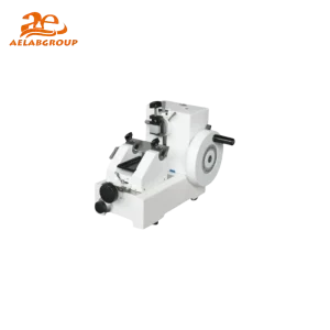
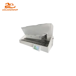
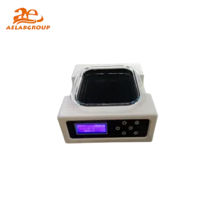
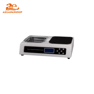
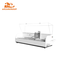
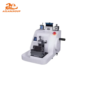
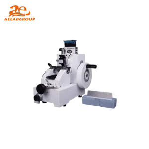
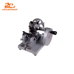
A Microtome is a precision instrument essential for creating thin tissue sections for microscopic analysis. Commonly used in histology, pathology, and biological research, microtomes ensure consistent section thickness, enabling accurate observation at the cellular and structural levels for diagnostics and advanced scientific studies.
A microtome is a laboratory device designed to slice samples into ultra-thin sections for microscopic examination. It consists of a sharp blade, specimen holder, and an advancing mechanism that moves the sample toward the blade with controlled precision. Microtomes are indispensable in histopathology, forensic science, and materials research, where uniform section thickness ensures reproducible analysis and imaging quality.
| Feature | Details |
|---|---|
| Section Thickness Range | 0.1 – 60 µm (depending on model) |
| Cutting Mode | Manual, semi-automatic, or fully automatic |
| Blade Type | Disposable steel, reusable glass, or diamond |
| Specimen Retraction | Automatic retraction to reduce wear on the blade |
| Specimen Size | Up to 50×60 mm (standard tissue block) |
| Ergonomics | Adjustable handwheel, anti-fatigue design, easy cleaning |
| Precision Control | Micrometer-driven or digital feed adjustment |
| Safety Features | Blade guards, locking mechanisms, emergency stop |
| Feature | Microtome | Cryostat | Ultramicrotome |
|---|---|---|---|
| Sample State | Paraffin-embedded | Frozen | Resin-embedded |
| Section Thickness | 1–60 µm | 5–30 µm | 50–100 nm |
| Use Case | Routine pathology | Surgical diagnostics | Electron microscopy |
| Automation Level | Manual to automatic | Temperature-controlled manual | Ultrahigh precision motorized |
| Cost | Moderate | High | Very High |
Q: What is a microtome used for?
A: A microtome is used to cut extremely thin tissue or material sections for microscopic examination, essential in pathology and research labs.
Q: How thin can a microtome cut?
A: Depending on the type, microtomes can produce sections from 0.1 µm to 60 µm thick, while ultramicrotomes can reach 50–100 nm for electron microscopy.
Q: What is the difference between a microtome and a cryostat?
A: A microtome cuts paraffin-embedded samples at room temperature, while a cryostat performs similar sectioning on frozen tissues within a cooled chamber.
Q: How do I maintain my microtome?
A: Regular cleaning, blade replacement, lubrication, and calibration are essential to ensure long-term accuracy and operator safety.
Looking for specific lab equipment? Fill out the form below, and our team will get back to you with detailed information and a personalized quote.