info@aelabgroup.com
QR Code 1
QR Code 2

Gel & Chemiluminescence Imaging Analysis System

Introduction

The Gel & Chemiluminescence Imaging Analysis System is essential for precise visualization, quantification, and documentation of nucleic acids and proteins. Designed for modern molecular biology and protein analysis, it delivers unmatched sensitivity, clarity, and reproducibility for gel electrophoresis, Western blots, and fluorescence assays. This guide covers its principles, advantages, and how to select the best system for your lab.


What Is a Gel & Chemiluminescence Imaging Analysis System?

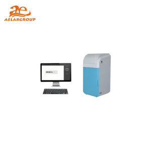
A Gel & Chemiluminescence Imaging Analysis System is a specialized device that captures and analyzes DNA, RNA, and protein samples after electrophoresis. Using fluorescence, colorimetric, or chemiluminescent detection, it provides digital, high-resolution images and quantitative band analysis for research and diagnostic applications.

  • Gel Documentation: Captures high-quality images of stained agarose or SDS-PAGE gels.
  • Chemiluminescence Detection: Visualizes protein bands using enzyme-driven light emission.
  • Quantitative Analysis: Measures signal intensity for accurate data comparison.

Key Components & Working Principles

  • High-Resolution Camera: CCD or CMOS sensors capture sharp, low-noise images, detecting faint signals.
  • Light Source: UV, blue, or white LED illumination for fluorescence and colorimetric visualization.
  • Emission Filters: Selective filters isolate specific wavelengths for accurate detection.
  • Imaging Software: Enables image capture, enhancement, densitometry, and quantitative analysis.

Technical Features and Specifications

Feature Specification
Camera Resolution 4–8 megapixels or higher for fine detail capture
Detection Sensitivity Low-picogram to femtogram detection limits
Light Sources UV, blue, and white LED illumination for multiple detection modes
Image Output Formats TIFF, JPEG, BMP for publication-ready data
Dynamic Range 3–5 orders of magnitude for quantitative accuracy
Software Compatibility Windows/Mac with advanced densitometry and reporting tools
Connectivity USB, Ethernet, or cloud-enabled data storage and sharing

Benefits

  • Detects low-abundance DNA, RNA, or proteins with high sensitivity.
  • Provides quantitative, reproducible data for publication and reporting.
  • Eliminates film-based imaging, reducing cost and chemical waste.
  • Delivers instant, real-time visualization and analysis.
  • Supports multiplex imaging with multiple excitation/detection channels.

Applications and Tests

🔬 Molecular Biology

  • DNA and RNA gel electrophoresis documentation
  • Fluorescent dye visualization (e.g., SYBR Safe, EtBr)
  • Gene expression validation and nucleic acid quantification

🧪 Clinical Diagnostics

  • Western blot chemiluminescence detection for biomarker studies
  • Quantification of diagnostic proteins and enzymes
  • Fluorescent antibody assays for immune analysis

🏭 Industrial & Food Testing

  • Protein stability and product quality testing
  • Verification of food allergens and additives
  • QC imaging for biopharmaceutical manufacturing

🌱 Environmental & Agricultural Labs

  • Detection of environmental DNA (eDNA) in soil and water
  • Protein and enzyme assays in plant biotechnology
  • Genetic diversity and molecular marker analysis

Chemiluminescence Imaging vs. Traditional Film

Criteria Chemiluminescence System X-ray Film
Sensitivity Higher detection of faint bands Lower sensitivity
Cost Over Time Lower—no film or chemicals Higher—requires film and reagents
Workflow Fast digital capture Manual and time-consuming
Quantification Accurate and reproducible Limited manual estimation
Reproducibility Excellent across runs Variable

Expert Tips for Choosing the Right Imaging System

  • Match camera resolution to your research needs—higher MP improves fine band clarity.
  • Opt for systems supporting multiplex detection for multi-target protein analysis.
  • Choose software with intuitive quantification, image overlay, and export functions.
  • Verify service availability, warranty terms, and local technical support.
  • Select systems with UV, blue, and white light options for maximum flexibility.

Maintenance and Best Practices

  • Clean imaging surfaces and lenses regularly to maintain clarity.
  • Calibrate camera and illumination systems as recommended.
  • Keep the device in a dust-free, temperature-controlled space.
  • Update firmware and imaging software to ensure optimal performance.
  • Handle gels and blots carefully to avoid contamination or smearing.

FAQ

Q: What is the main advantage of a chemiluminescence imaging system over X-ray film?
A: Digital imaging provides higher sensitivity, better quantification, and eliminates film processing, saving time and cost.

Q: Can the same system be used for both DNA gels and Western blots?
A: Yes, hybrid systems support gel documentation, fluorescence, and chemiluminescence modes in one platform.

Q: How often should the imaging system be calibrated?
A: Calibration should follow manufacturer recommendations—typically every 6 to 12 months or after major software updates.

Q: What causes faint or blurry chemiluminescence bands?
A: Check the freshness of substrates, ensure even illumination, and optimize exposure settings and antibody concentrations.

Looking for specific lab equipment? Fill out the form below, and our team will get back to you with detailed information and a personalized quote.